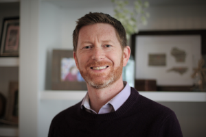
In Vivo Imaging, Muscle Neuromechanics, and Rehabilitation for OA Prevention – December 15, 2021
December 15, 2021
Speaker:
Associate Professor, Joint Department of Biomedical Engineering
Faculty Affiliate, Human Movement Science Curriculum
Faculty Affiliate, Thurston Arthritis Research Center
Director, Applied Biomechanics Laboratory
University of North Carolina at Chapel Hill
North Carolina State University
Dr. Franz received his B.S. (2004) and M.S. (2006) degrees in Engineering Mechanics from Virginia Tech and, after serving as a staff scientist in PM&R at the University of Virginia, received his Ph.D. (2012) in Integrative Physiology from the University of Colorado, Boulder. He then completed an NIH Post-Doctoral Fellowship in the Department of Mechanical Engineering at the University of Wisconsin-Madison. In 2015, Dr. Franz joined the Joint Department of Biomedical Engineering at the University of North Carolina at Chapel Hill and North Carolina State University and is now an Associate Professor and Director of the UNC Applied Biomechanics Laboratory. He currently serves as Principal Investigator or Co-Investigator on multiple NIH-funded research projects, all predominantly focused on rehabilitation engineering strategies to mitigate age- and disease-related mobility impairment and falls risk.
Lunch & Learn Recording & Transcript
Disclaimer:
The content displayed in this transcript is the intellectual property of Jason Franz. You may not reuse, republish, or reprint such content without written consent. The contents are those of the author(s) and do not necessarily represent the official views of, nor an endorsement, by OA Action Alliance or CDC/HHS. This transcript was automatically generated in Zoom, and edited for clarity; however, the OAAA cannot guarantee there are no mistakes or errors.
December 15, 2021
Title: In Vivo Imaging, Muscle Neuromechanics, and Rehabilitation for OA Prevention
Presenter: Jason R. Franz, Ph.D.
Associate Professor, Joint Department of Biomedical Engineering
Faculty Affiliate, Human Movement Science Curriculum
Faculty Affiliate, Thurston Arthritis Research Center
Director, Applied Biomechanics Laboratory
University of North Carolina at Chapel Hill
North Carolina State University
INTRODUCTION
(Kirsten Ambrose) Hello, and welcome to Osteoarthritis Action Alliance Lunch & Learn webinar for December 15, 2021. Thank you for joining us this month, our presenter today is Jason Franz, Dr. Franz received his Bachelor of Science and has Master of Science degrees in Engineering Mechanics from Virginia tech, and after serving as a staff scientist in physical medicine and rehabilitation at the University of Virginia received his PhD in 2012 in integrative physiology from the University of Colorado Boulder. He then completed a postdoctoral fellowship and the Department of mechanical engineering at the University of Wisconsin Madison. Franz joined the joint department of biomedical engineering at the University of North Carolina Chapel Hill and North Carolina State University and is now an associate professor and director of the UNC Applied Biomechanics Laboratory. He currently serves as principal investigator or co-investigator on multiple funded research projects all predominantly focused on rehabilitation engineering strategies to mitigate age and disease related mobility impairment and falls risk. Dr Franz’s talk today is titled “In Vivo Imaging Muscle Neuromechanics, and Rehabilitation for OA prevention.” Welcome Dr. Franz.
PRESENTATION
(Dr. Jason Franz) Thank you for the invitation and the introduction, can you hear me okay? Does everyone have their lunch ready? I’ve spent the last 15 minutes scarfing mine down, but I welcome you, you can totally eat, while I talk I won’t be interrupted, I promise. I also noted some familiar faces in the participant list so it’s nice to see some friends here, and some of the content, if not, you know much of the content here might be stuff you’ve seen before, but the more perhaps we think about these ideas, there might be new opportunities for collaboration. But, as you heard in that introduction I direct the Applied Biomechanics Lab, which is in our joint Department of Biomedical Engineering UNC and NC State fully joint both undergraduate and graduate levels as well as our faculty and the goal of my lab is twofold. One, I want to collaborate with our students and our faculty collaborators to understand the mechanisms involved in losing one’s independence in their community as they get older, and I want to leverage that newfound understanding to engineer new strategies, devices, and approaches to mitigate that loss of independent mobility. This mission is very familiar to many of us, because you know many of us have had or are currently having older relatives or older neighbors that we’ve watched suffer from declines in physical function and mobility and an increased prevalence of faults, this one here happens to be my grandfather and like many of your older relatives or neighbors I watched him go from just being the strongest most physically fit and capable person I knew to becoming physically weaker, becoming frail, falling more often, and certainly suffering from the degenerative impact of osteoarthritis.
So that’s the motivation for all of the work that we do and with our short time together today I’d like to follow a pathway that we think is taking place on a step-to-step basis that we think ultimately contributes to the onset and development of osteoarthritis. This is a relatively new area of research in our lab over the last couple years. And what I hope to do is to introduce some new things here about the role of muscle itself in this pathway. You know some of this might be biomechanical some of this might be inflammatory but ultimately, we propose that those two things are actually highly interconnected. We also think that some of this pathway might be common to both post-traumatic osteoarthritis following knee joint injury earlier in life and idiopathic osteoarthritis later in life and I’d like to sort of characterize our work in both of those areas. Much of our work here at UNC is performed in collaboration with folks at the UNC Motion Science Institute. Some of those have actually performed or come and given this this particular talk at this lunch and learn and they for years have studied ACL injury and reconstruction as a model for the development of osteoarthritis specifically post traumatic away. And there’s well documented strength and activation muscle deficits in the quadriceps muscles following ACL reconstruction. And those are at least accompanied by altered knee joint biomechanics during walking considered relevant to osteoarthritis. But because you know that the quadriceps muscles themselves are by far the largest contributor to joint contact forces during walking.
Those changes are thought to alter cartilage loading patterns to magnitudes and with locations that aren’t protected by prior loading experience and there’s a lot about this. We’re calling it a neuromechanical cascade that we simply don’t understand, but you know, allow me as you eat your lunch to focus on what I think are two major opportunities are gaps in our understanding here.
The first is that deficits in quadriceps force generation and subsequent changes in gate by mechanics, they’re not simply governed by weakness and neural changes inhibition. The actual mechanics of the muscles involved here really aren’t well understood even in healthy and uninjured adults. And the second is that study is trying to associate altered gate biomechanics with altered cartilage loading. They’re purely observational, it makes it nearly impossible to establish cause and effect relations between factors that may predispose to us to arthritis and so, if we can overcome those, you know, even some of those challenges, we think we might be able to identify new or more effective translational opportunities for prevention.
Now there’s three skills that our lab brings to the table to address these gaps with our collaborators, so the first is ultrasound imaging looking under the hood at the muscles powering locomotion real time, the second is gate biofeedback, a super simple idea, providing you with information about what you’re doing and the hopes to steer that in a different direction. And then, lastly, if we must go scout for a model because we cannot physically measure the thing we’re interested in, maybe we can use the computational power of models to do that. And our original work here set out to adjust two main questions, the first is: how does joint injury impact quad muscle mechanics underlying gate biomechanics? And the second is can we manipulate joint loading and tissue biochemistry to optimize rehab and prevent osteoarthritis.
And we know that quad dysfunction after ACL reconstruction is accompanied during walking by reduced extensive moment as you can see here on your left. And deflection excursion which you can see, on your right. Both of these changes are evidence of quadriceps avoidance during walking, but this is clearly an incomplete story because, you know, conventional resistance and training, they can restore muscle strength, but those interventions do little to actually mitigate these gate changes. And like I said In Vivo Imaging provides a way to look under the hood discover how these muscles are actually operating to produce the new moments and emerging joint contact forces considered so relevant to OA.
And so what you see here in this plot is length change of the entire muscle tendon unit, so this is from proximal to distal bony attachment. And we focus on this shaded region, the weight acceptance phase, and here the quad muscles are generating forced activity, resistant selection, and muscle tendon unit lengthening. However, if we image the muscle directly we actually find something really surprising. As the knee is flexing and the entire muscle tendon unit is lengthening the quad muscles themselves are actually receiving sufficient activation to shorten as the joint is being loaded. And what that implies is that tendon lengthening, not muscle lengthening is more responsible than we’ve anticipated, governing the forces that are generated by the quadriceps during limb loading and walking, at least in uninjured controls.
Which brings me here, so what happens following ACL injury and reconstruction. Well, let’s read take those same measurements during walking in individual six to 12 months after reconstructive surgery. And as a reminder these individuals walk with big reductions in the extensive moments generated by these muscles, the forces are lower, and here we see the quadriceps muscle action in the uninjured group, so the uninsured contralateral limb is not really different or indistinguishable from uninjured controls. But we see fundamentally different behavior on reconstructed limb. Only there do we actually see significant muscle lengthening during limb loading, which is even more surprising, you know, given that this is going through much less knee fluctuation than uninjured controls So what does this mean well, we have two possible explanations, both of which we think are relevant strategies that might optimize knee loading following joint injury and ACL reconstruction.
The first is potentially because of insufficient muscle force, the muscles themselves may be either succumbing to the demands of walking or actually shifting to ease centric muscle action as a compensatory strategy and then, the second is that these individuals may operate their muscles, in a way to avoid excessive tendon lengthening something especially relevant in these subjects who all received bone patella bone grafts. But what we’re getting more and more excited about and interested in is there might be significant inflammatory consequences associated with early signs of cartilage and bone degeneration. As a first takeaway of this talk, really these data suggest that the quadriceps muscles themselves may serve as a marker of recovery or target for intervention. To reduce the risk of post traumatic OA for individuals following ACL reconstruction and those inflammatory consequences there, that’s all-animal data showing that lengthening contractions might change the inflammatory state of the joint.
Alright, so our second key question, to start with here: can we manipulate joint loading and tissue biochemistry to prevent OA? And again, here we’re trying to move beyond merely observing differences between groups. We know that people following ACL reconstruction walking away, we would suspect based on this curve, at least to under load the articulate cartilage compared to uninjured controls. And this idea of under loading has implications for cartilage tissue integrity. It was about three years ago we show that individuals with less limb loading measured here by smaller peak vertical ground reaction forces on the y axis. They actually exhibit larger changes in serum cartilage alert matrix protein or come on the on the X axis after just 20 minutes of walking. What that means, it suggests more cartilage breakdown in individuals who exhibit less limb loading during walking.
Now, as I mentioned earlier, we’re really interested in biofeedback both as an experimental tool and, ultimately, as a means to enhance our optimized rehab. And in the study, here we set out to determine if augmenting limb loading during early stance and walking using biofeedback could also augment that biochemical response relative relevant to cartilage tissue integrity. So here on different days separated by about a week subjects are going to walk while watching a video screen showing their instantaneous peak vertical ground reaction force that we measure from a treadmill. They will be measured on a step-by-step basis and they’re going to see a target line that’s going to represent usual loading, lower than usual loading, and higher than usual loading, and we’re going to measure the change in serum calm following 20 minutes of walking with each of these biofeedback conditions. The key outcome from this study is here, we found that prescribing higher than usual loading with biofeedback actually decreases the change in CRM COM suggesting that we can augment joint loading and tissue biochemistry using biofeedback. The challenge here is that peak vertical ground reaction force is sort of a limb level indirect surrogate for knee joint loading. So, we’re interested in knowing you know, can we more directly target the knee while simultaneously in our lab we’ve been performing more direct real time estimates of extensive moments on a step-by-step basis. To do this, we’ve been using biofeedback and all of our fancy equipment in the lab and those estimates agree pretty well with those you would get from inverse dynamics. How the laboratory typically goes about calculating these in a really time intensive way. And here, what we’re going to do is we’re going to encourage visual by using visual biofeedback to encourage subjects to walk with larger than normal techniques, and sensor moments with the dots shown in green and walk with smaller than normal techniques denser moments with sort of the dots shown in blue. In doing so, you can see here that we can actually manipulate the denser moments on a step-by -step basis to prescribe values here, showing on the left. And, and these changes are accompanied by changes in knee FLEX and excursion, in other words, subjects are encouraged with my feedback to overcome that quadriceps avoidance strategy underlying stiffening gave up to contribute to altered cartilage loading.
But again, we’re trying to dive deeper, we’re trying to move beyond just observing things so let’s not stop there, because I think we can do better in terms of establishing cause and effect relations between altered gate biomechanics and cartilage tissue loaded. So, what we do here, and when we you know in my lab when we can’t actually measure something directly, we leverage some of the recent developments in in musculoskeletal modeling. And, in our case we’re going to use a simulation pipeline called komack. This is…I’m just going to read it here because it’s very long concurrent optimization of muscle activations and kinematics. And what’s cool here is that we can actually simulate combinations of muscle activation weights and soft tissue properties to account for individual variation between people and you know functional anatomical variability mechanics or tissues super powerful tool. So, we’re doing a number of things here.
Let’s drive that model using measured whole body motion capture and ground reaction forces from our treadmill and subjects walk using that real time extensive moment biofeedback. And just to show you here, these are some of the preliminary data that we have so far, a student got group average data as of this week which will be ready for abstracts this season. All the data points presented here change relative to normal walking without biofeedback and on the left, we can see change anterior typical translation. And on the right, we can see change in peak force transmitted through the ACL and what I want you to get across from both of these is that these outcomes within the joint are sensitive to prescribe changes and extensive moment. I don’t want you to take away the same thing from this, we can actually estimate changes in cartilage contact for us which I’ve separated here into force in the medial to be a femoral plateau is the closed symbols and on the lateral to the ephemeral club toes the open symbol. And again, just showing that modifying gate with biofeedback has an emergent changes, all the way down to the magnitude of forces experienced by the particular cartilage within the knee is a really powerful paradigm. And those data suggest that we can use this type of approach to augment, or perhaps optimize mechanical loading and the need to preserve and restore cartilage tissue integrity in hopes of preventing the development or maybe slowing the progression of OA. The obvious challenge, there is translational feasibility and I’ll simply say you know, there are really terrific advances in the use of wearable sensors and machine learning that may eventually be able to overcome those challenges and allow for some scalability and feasibility in the hands of clinicians.
So where do we go from here, allow me to close here just with a new direction or application of all of this work, as we merge our years of work. Looking at the biomechanics of movement in older adults with our more recent contributions to understanding the neuro mechanical pathway to osteoarthritis and the involvement between the interaction between muscle action and biomechanics and the inflammatory state within the joint being so with new funding, we were very gracious and very happy to receive from UNC. We’re in the process of testing the hypothesis, you see here in older adults with idiopathic osteoarthritis, and that is in older adults with OA. The hypothesis is: Do the quadricep muscles succumb to the demands of lower extremity loading with aberrant lengthening action with every single step? Because if they do, this has the potential to increase inflammation and cartilage contact forces. So, we’re just getting ready to start subject recruitment here, we should know more about this, this time next year, and we can share it at another lunch.
So, with that, I really want to emphasize all the wonderful contributions of our group and our collaborators. I’ve put a big black box around the PhD student who’s most heavily involved in this work, Mandy, a fourth year PhD student in our group. And again, I really appreciate you sharing your lunch with me and giving me the chance to share a little bit about our work and I’d be really pleased to take any questions you might have.
QUESTION AND ANSWER
(Kirsten Ambrose) Thank you Jason! I think this space in between the original joint injury to end the development of post traumatic is fascinating, and we still have so much to learn. Great presentation, giving us a little bit more insight. As a reminder, just like Jason said, we do have time for questions. You can type your questions in the chat box and also in a few minutes you’ll see a poll pop up on your screen, with some survey questions so we’d be appreciative if you’d answer those before you leave the webinar.
QUESTION: How would you implement biofeedback modalities to augment joint loading and biochemistry in the real world?
ANSWER: Dr. Jason Franz: Yeah, so, the funny thing is right now right all this work takes you know a $150,000 treadmill, so it gets totally ridiculous to think that there would be access, accessibility, and equity in the delivery of some of these technologies to physical therapy clinics. But we have ongoing work that is trying to see if we can Whittle all of this laboratory equipment down to a small set of wearable sensors. Whether those are sensors that you might place on the on the limbs, or maybe planner pressure data, can we replicate this laboratory system in a really simple take home take to the clinic type approach. And I’ve been really inspired by some of the developments in machine learning and all the great investigators using wearable sensor work I think it’s totally feasible and so that is sort of my vision for where all that all that would go.
(Kirsten Ambrose) Thank you, I think it’s definitely important to make this relatable in a real-world setting. It’s great to do the laboratory work that’s necessary, but then, how do we make it…how do we translate that.
QUESTION: Thank you. Very, very interesting! How strongly did deflection excursion angle correlated with the km and the other parameters difficult to assess?
ANSWER: Dr. Jason Franz: That’s a good question. So essentially saying you know, to get an extensive moment, not only do you need motion capture you also need forces, and I think what you might be suggesting is you know, is there a way to get to a surrogate like deflection angle, the excursion reflection angle that we could assess in the lab. These things do change in the same direction, so, for example, if we prescribe an increase in the extensive moment yourself, that’s also a company by an increase in FLEX and excursion so that’s good news. The magnitude of change can be really different and then what the big challenges is, I think that the way those two things relate to one another, can differ by individual and different populations. So even if you had a nice collection excursion which you might be able to just get with a camera or your phone even, if you optimize that for one person, it’s hard to know how it might translate to the next person or if you come up with some sort of…if you’ve made some clinical decisions on treatment, based on the relationship that you found, can you then go ahead and apply that clinical decision to other people that exhibit the same amount of new FLEX excursion? I don’t know, and I’d certainly want to test it. But I totally understand where you’re coming from, and it speaks to this idea of trying to get the science out of the lab, which is something we think about quite a bit.
QUESTION: Great presentation and very interesting, thank you for sharing your work. Did you measure muscle length from an image taken during walking or at rest?
ANSWER: Dr. Jason Franz: In the study that we’ve published in young adult controls and injured controls and then in the study that’s in review, in the individuals following ACL reconstruction those we only captured images during walking. But we’ve sensed come back and we’re doing a follow up study in a lot of people with ACL reconstruction, participating this biofeedback, and doing a lot of modeling and there we’re getting static images of the quads at various knee angles. So, we’re trying to. We’re trying to completely characterize the operating landscape of the quads from isolated contractions and rest and then, when we get our walking data, the vision is then we can sort of layer on where was your muscle during walking and how does that relate back to that some of those static measurements. And this is because some clinics might have access to ultrasound, but maybe they don’t have the capacity to measure these things, while the people are moving so again, it might be nice to define ways to make it easy on clinicians if things correlate.
QUESTION: Thank you for sharing your expertise with us, are there any other known mechanisms, other than increase loading that will help to prevent post traumatic knee arthritis my understanding is that controlling inflammation is helpful?
ANSWER: Dr. Jason Franz: I am going to plead the fifth sherry I am new to this space, and I think it would be overstepping my area of expertise to talk about specific treatment, my understanding is the same as yours that biomechanics are important and inflammation is important and lessening the inflammation, whether through therapeutics or other means is helpful and preventing osteoarthritis. I think what we add here is that those two things might be sort of fundamentally connected that the loading the muscles actual behavior in response to or to generate that loading and the result and inflammation might be this really highly interconnected thing. So, to answer there, the idea of controlling inflammation you can do it directly through therapeutics but if the source of some of that inflammation is the behavior the muscle itself then you might have to go to the biomechanics first and indirectly control inflammation. So, I think it gets complicated. Thank you for your question.
QUESTION: Thanks for your talk. Your initial data seem to show that more AP translation came with higher loading, but was that increase good or was it considered exaggerated?
ANSWER: Dr. Jason Franz: Yeah so the data that I’ve been able to present so far has been on single subjects and we haven’t really put too much emphasis on it and that’s why, you know, you identified a specific result, and what I did when I present it was like look at how these things change, right, because I don’t want to put too much emphasis on a specific directionality or the specific magnitudes. I am waiting for the morning, which could be even be like tomorrow, when Mandy who I mentioned is at the very end of the simulations that are incredibly difficult, they’ve taken about 18 months to work through, and I think it’s any day now where will actually know does higher loading change, you know it’s your post your translation in this direction, does it increase or decrease ACL force does it change to be a femoral cloud so contact forces and in this direction by how much so stay tuned.
CLOSING REMARKS
(Kirsten Ambrose) Wonderful! Well, thank you so much, and we are at the end of our 30 minutes. If anyone is typing a question, as I talk feel free to keep typing and we’ll answer it. The poll just popped up on your screen and I just want to thank everyone for joining us today, please join us in next month for our January 19 Lunch and Learn featuring our good friend Ellen Schneider who will present, “Expanding the Arthritis Appropriate Evidence-Based Intervention Menu,” so we look forward to sharing that with you, and in the meantime thanks again Jason and to everyone, we hope you have a wonderful holiday season and we’ll see you next month.
COST
Free

